Question about horn shape of abalone pearls

Steve, I think that more than nacre secretion away from the mantle, in the abalon case it?s because of the "alternant" (hope this is the word) nacre deposition system, it is the conchiolin the one that is been secreted away from the mantle, the nacre formed is just an special arrange of the calcium in the inmediate area. Sorry for my english, I can say it in spanish for better explanation if it?s not understood.
On the other hand, I have the abalone shells. I?ll upload the pictures of them for you to chose a couple (they 9 or 10).
 
Tango,

Many thanks. The dilemma represented by this thread is a very straightforward one (albeit pivotal to potential issues we are facing with Nautilus) and I look forward to presenting your specimens to prominent marine biologists for study.
 
One of the images
zoomaconcha.jpg
 
Assuming the question is for me, I?m not shure about the origin of the nacar piece in your picture. The image is not very clear but it may be formed because of a different reason from the ones on my shells.

In that area, the gonadal area, there isn?t a peritoneum that holds the organs and separates them from the shell. There is also a small mantle tissue just outside this area and if you press with your nail on the limit between the external membrane and the shell it opens easily.

So there you have an explanation for it, obiously it?s just an idea.
 
I've been interested in reading the posts regarding how an abalone tusk pearl is actually formed. It seems to me that my large broken pearl might be of some interest.
Patricia's post above on 'Natural Abalone Pearls' along with her link to prior posts of her two-piece 'tusk' pearl belongs here.

Patricia's pearl is indeed interesting. I have rotated her posted image and extracted the gonadal activity from Tango's shell, side-by-side below. This seems to support the gonad-hepatopancreas interface theory, exterior and interior sections unsuccessfully fused in this case.

I have added to the image our 52-carat oddity, purportedly from Nautilus, involving the joining (in this case partial, as with Patricia's ab) of two parts. As with gonadal abalone pearls, a biological process beyond the mollusk's mantle may be involved.
 

Attachments

  • Ab-Naut.jpg
    Ab-Naut.jpg
    74.3 KB · Views: 38
... a biological process beyond the mollusk's mantle may be involved.

The divers brought me an interesting specimen.

While not abalone, I thought I'd post these pictures of a horn and star formation from a Purple Hinged Rock Scallop (Crassadoma gigantea). This scallop shares the family Pectiniidae, but is often overlooked. Perhaps it's because of the sedentary nature of the adult, as opposed to the mobility of other scallops.

Either valve had a horn shape alternately embedded within the adductor muscle. Each presented with evidence of borer predation, likely Polydora. One horn didn't arrive with the specimen as it was probably sectioned when shucked. The other broke free quite easily. They are semi-hollow and somewhat chambered internally. I removed approximately 1.5 grams of necrotic tissue from both, but found no live parasites, other than smaller ones nearby and tiny ones near the edges of the valves.

The image shows the horn made up mainly of conchiolin, but hardened by minor amounts of calcium carbonate.

The other blister, the star shape presented with no evidence of parasites, but as "filling the space" between the adductor and gonad, much like my description earlier in this thread about squeezing puddy.

Unfortunately, this specimen was intended to be eaten and not prepared for microscopy. The epithelial cells taken from tissue sections were not stabilized in a timely manner and as such did not react properly with stain.

However, this still is a very good sample, because it demonstrates both horn-like parasitical cysts and auto-immune defensive mechanisms within the same specimen.
 

Attachments

  • scallop05.jpg
    scallop05.jpg
    25.7 KB · Views: 41
  • scallop02.jpg
    scallop02.jpg
    19.3 KB · Views: 50
  • scallop06.jpg
    scallop06.jpg
    22.3 KB · Views: 35
  • scallop04.jpg
    scallop04.jpg
    23.9 KB · Views: 38
  • scallop01.jpg
    scallop01.jpg
    20.7 KB · Views: 39
  • scallop anatomy.jpg
    scallop anatomy.jpg
    11.9 KB · Views: 37
An interesting specimen for sure.

However, the dark conchiolin deposits do not appear to be horn shaped to me, rather as amorphous lumps. There do seem to be somewhat horn-like porcelaneous shapes in the shell vicinity. Is that the observed phenomenon?

Cutting to the chase, do these observations suggest any conclusions regarding the formation of nacreous gonadal abalone pearls?
 
It suggests infestation by parasites may give rise to autoimmune disease in the gonads of molluscs.
Thereby causing the gonad epithelial tissue to secrete conchiolin/nacre in an attempt to recreate itself posthumously?
 
There do seem to be somewhat horn-like porcelaneous shapes in the shell vicinity. Is that the observed phenomenon?

Naturally, there are several active epithelial sites near the gonads in all molluscs and of course, it's the highly compatible tissue/nucleus grafting site in round pearl culture. So nearby, things like extra layers, wider margins, varied structures etc. are common place.

Where anomolies occur, the absence of trauma almost always points to the blood.

I think it?s some kind of a chain reaction produced mostly by a bacteria or protozoo.

Again, most likely transmitted via the blood.

In that area, the gonadal area, there isn?t a peritoneum that holds the organs and separates them from the shell. There is also a small mantle tissue just outside this area and if you press with your nail on the limit between the external membrane and the shell it opens easily.

Hence a super-sensitive organ is super-vulnerable and an ideal place to detect early phases or record long term histories of disease.

So lets assume my theory holds true for a moment. The next questions would be A- Septic or aseptic at the affected site? and B- (in the case of the topical abalone) Is the disease onset in the gonad itself, or from the surrounding tissue?

To that, my answers would be, any combination is possible, but ... to go out on a limb ... suggest aseptic inflammation of the surrounding tissue (closely resembling mantle tissue). The organ continues to function, but at a lesser rate within the formation as the animal and pearl grows.
 
Last edited:
So, chronic inflammation initiated by the parasitic infestation, whether or not the infestation is walled off...no wait, you're suggesting the infestation is caused by something way smaller than usual, capable of traveling through the bloodstream but not clogging it up, the way a virus can?

Forgive the ignorance, just trying to follow along.

I'm thinking along the lines of where the immune system is fooled into attacking otherwise healthy tissue. I also suggest there could be both nuclear and anti-nuclear factors among individuals as well. Nuclear being encapsulation of cells and anti-nuclear being destruction of cells.

Shells are vulnerable to trauma, so wide areas of the mollusc must go into repair mode, immediately, at any time. This is especially true with the tissues around the gonads, clearly from an evolutionary and survival standpoint. Anything with weak nads would have died out eons ago.
 
Protozoos can use the blood stream to travel too Lisa, but only when the infestation is big, the same for bacteria, septicemy.
The haliotis circulatory system is open, there for, it?s easier to be infected than ours.
By antinuclear you mean apoptosis Dave?
 
Last edited:
By antinuclear you mean apoptosis Dave?

Perhaps, but not entirely. Apoptosis is the programmed death of cells which occurs naturally in multicelled organisms. In cases where apoptosis is premature or accelerated, then there is evidence of antinuclear behavior.

Hemocyte phagocytosis represents the main process of the entire cell defense system and is constituted by different phases including recognition, adhesion, ingestion, destruction and elimination of foreign cells. Mollusc hemocytes also have important
functions in transport of nutrients, wound/shell repair, and removal of catabolic products or pollutants.

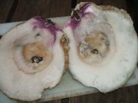
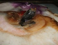
The greater portion of pearl nucleations fail when excessive counts of hemocytes surround the graft. Usually by excessive bleeding. Sometimes grafts fail when surrounded by gametes, as the gonad is damaged during the operation.

This is from Roch, 1999: (I've highlighted relevent points)

HUMORAL DEFENSE SYSTEM

Invertebrates possess aspecific and innate immune mechanisms, and they are lacking
in immune memory following the first encounter with a pathogen
. Molluscs have
humoral and cellular immunity, and the humoral system is constituted by lysosomal
enzymes, agglutinins, lectins and antimicrobial peptides. Nevertheless, cellular immunity
seems to perform the main role in shellfish immune processes.

Lysosomal enzymes (b-glucuronidase, acid and alkaline phosphatase, lipase, aminopeptidase and lysozime)
are comprised especially in granular hemocyte lysosomes
and their release into serum depends on cell degranulation during phagocytosis
(Pipe, 1990). Lysosomal enzymes also possess digestive function as indicated by their
abundance in the stylus and digestive gland. In fact, digestive and defense functions
could act contemporaneously because filtered bacteria represent nourishment for
marine bivalves, so enzymes present in the digestive tract hydrolyze microorganisms
performing both functions at the same time.
Agglutinins have been found in many
mollusc tissues and these glycoproteins act as opsonins against different types of
erythrocytes (hemagglutinins) and other cells such as bacteria, protozoa and algae
(Chu, 1988). Lectins represent agglutinin-like molecules and are present in the hemolymph
and in the hemocyte membrane; they have a specific opsonizing function in
mollusc defense mechanisms such as hemocyte aggregation and foreign cell agglutination.
In particular, lectins are specific for hemocyte and bacteria glycoconjugates, so
promoting recognition of foreign cells. However, the biological significance of the
different carbohydrate specificities is not yet known, in fact most invertebrate lectins
are heterogeneous and may also bind other ligands (Tunkijjanukij et al., 1998).
Antimicrobial peptides have been discovered in hemolymph and mussel hemocytes,
and have been classified as defensins, mytilins, myticins and mytimycin. These compounds
act on a rather broad spectrum of microbial organisms and their activity has
been shown outside hemocyte or in phagocytosed bacteria
(Mitta et al., 2000).

To to summarize my point, when infections occur, otherwise healthy cells can be collaterally affected, whether by the pathogens themselves or incidentally by other blood components.
 
Interesting hypothesis.
There is the point never the less, of the formation of this inflamation outside the circulatory system, next to the shell, where aren?t any of this lyzosomal enzymes, lectins, etc. So this phenomenon would be explain only in the intraperitoneum space.
 
Back
Top