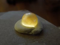
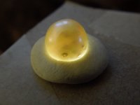
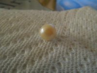
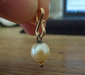
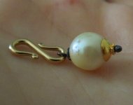
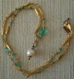
You know what? The more I look at this pearl, the more I think we're mistaken.
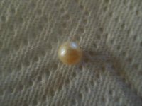
I candled a few 20+ year old undrilled akoyas I had stashed away.
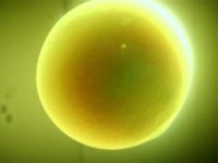
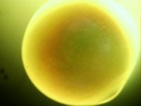
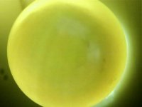
Other than the blemishes and gas inclusions, I'm seeing similar structures below the surface, but no deeper inclusions. There is a slight variation in the geometry of the stippled surface, but you can clearly see it's just below the surface.
I noticed you've since fashioned the pearl into a finished piece, but I might have been inclined to look down the holes a little closer.
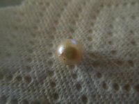
One other concern, the layers of the nuclei. Look closely at the third image (note: edited from "first"), (albeit slight) the appears to be evidence of layering in the nuclei. In the last of the akoya images, they are seen
I have posted a few images of nuclei from American river mussels. Two species, washboard mussel with dark lines and pigtoe mussel with lighter lines.


One other point I should make. A single axis view is incomplete data. To be objective, it's important we obtain views from X, Y, and Z axis, systematically.
Here is the reason why, this image is the same washboard mussel in the first image of nuclei, just along a different axis. You can see the marked difference, that must be interpreted according to the other axis (plural).

Examine all of these images closely for a few minutes, then go back to the first page.
What do your eyes think?

