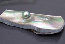
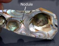
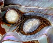
This fifth one is the one that really got my attention and caused me to realize what the nature (I think) of these objects are. It's overall size is about 50mm X 32mm. The center node is about 4.6mm X 4.5 X 4mm. The little one to the right of the center is about 2.8mm X 2.5mm X 1mm.
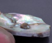
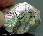
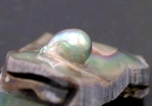
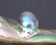
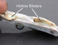
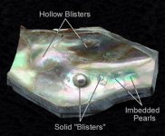
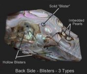
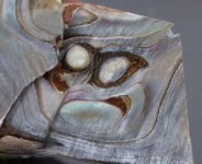
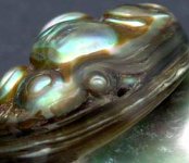
If you will note, the first photo shows three different types of protuberances. you may also note what all three (?) of these blister types look like from the partly ground back. The hollow back blisters are the result of toredo clams boring into the shell, the cause of by far the great majority of the blisters in the abalones I have. The center nodule and the small one to its' right show no discernable evidence of their existence from the back. The two low dome blisters are a different story, however. When I examined them closely, I saw on the back that directly under each of them I had nearly bisected two objects surrounded by conchiolin. Under magnification I could see that their structure consisted of coencentric rings of what appears to be nacre. The next two photos are close-ups of these. For the second one, the most enlarged, I raised the contrast to make the rings more visible. In the more oval one the cut seems to have gone to almost the exact center of the rings where there appears to be a very tiny, round knob of about .8mm diameter.
In Berry Blister #1 I mentioned that the way it had been ground and finished may show a clue to it's origin. The last picture is of one side of #1 showing one of the nodules ground part way through. Though not ground as far as the two in the fifth one, it too shows what I think is most likely a coencentric layered structure that goes down to a small center.
What I think that this means is that these solid "blisters" began life as free pearls grown in the mantle of the abalone. Somehow they were dislodged from the mantle and came to rest against the shell. They remained trapped there long enough for the abalone, perhaps feeling them as an irritant, to cement them to the shell with conchiolin and nacre and then to continue coating them with layers of same. Two of them, #4 and #5, appear to have the same or a smaller diameter at the base where they intersect the shell as they do at midpoint. In pictures I have seen of small free pearls being removed from the mantle they look as though they hold a rather tenuous connection to this mantle. Most of these "pearls" that I have, I have found up inside the lip of the shell.
Historically. some very valuable free pearls have been uncovered in salt water bivalves at the site of a large blister. These shells, having been subsequently peeled down by a pearl pealer, eventually revealed their prize. I am beginning to think that most, or perhaps all of the solid "Berry Blisters" and single nodules I have shown here are the result of free pearls or clusters of free pearls having been ejected or somehow dislodged from the mantle and subsequently then attached to the inside of the shell and coated with layers of conchiolin and nacre until they became part of it's structure, some of them perhaps being so covered that they eventually became a hard-to-discern part of the body of the shell.
At this point I am considering attempting to dislodge one or both of the suspected free pearls from example #5 to see if the hypothesis proves out. I have found a few other examples and possible examples of this type of anomaly in some more shells. The new ones that show on the surface are, so far, very small and the "possible" ones are deep in the interior of the shell, if that is indeed what they are, and will have to be exposed. Doing so will take some time, with all the other work that I have to do, so I don't know how long it will be before I am able to do this. Meanwhile, I have some other, very different, solid blisters that I will try to post in this thread. I am very interested in hearing what other forum members think about all this.

Marc
http://flyrodjewelry.com/home.html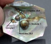

") Non-destructive test ...
Non-destructive test ...