Lagoon Island Pearls
Well-known member
- Joined
- Dec 8, 2009
- Messages
- 2,152
I've been back in the field collecting specimens. I set out this year to revisit a theory on the incidence of pearl formation related to heart disease.
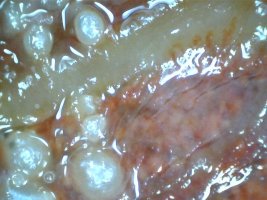
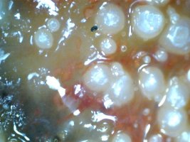
In almost every specimen found to contain pearls, a great percentage were found within <50% of the radius from the center of the anatomy. This suggests heart involvement. Mussels don't have hearts as a single organ, insomuch as laddered pericardium. From the heart, capillary ducts and valves interconnect the vascular system which also supplies the reproductive systems through a series of gonoducts. Together with skin, muscle and fascia theses form the pallial mantle.
I've started looking much closer at the heart in every case now and it seems the incidence higher than previously anticipated.
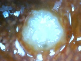
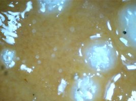
Microscopic analysis is revealing some interesting things. It was exciting to find pearls with visible sacs attached to the linings of the heart. No only is this revealing, it's revolutionary! Why? It is because we are witnessing the birth of pearls.
While other causes for onset are known, this one is different because these pearls are sterile and rather than having a nucleus itself, they are the nucleus of the lesions themselves.
Now that I have sacs, I can stain for protease. A protease is an enzyme that catalyzes proteolysis, breaking down proteins into minor polypeptides or single amino acids, thus stimulating the formation of new protein products. That's going to take time because my surgery is crude, but I'm searching new micro-biopsy tooling.
Stenosis is the obvious result, but not the cause which is yet to be determined. Mollusks have an open circulatory system, therefore this is not a valve issue. Pearls appear to form at any position along the vessels. Many will irrupt from the sac, thus becoming lodged elsewhere within the circulatory system, sometimes restricting blood (hemolymph) flow.
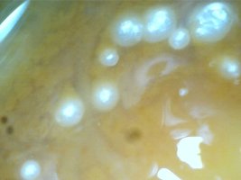
I've also noticed several pearls attached near the hinge and always on the right shell. Another clear sign of natural pearls from maladies at or near the heart.
In almost every specimen found to contain pearls, a great percentage were found within <50% of the radius from the center of the anatomy. This suggests heart involvement. Mussels don't have hearts as a single organ, insomuch as laddered pericardium. From the heart, capillary ducts and valves interconnect the vascular system which also supplies the reproductive systems through a series of gonoducts. Together with skin, muscle and fascia theses form the pallial mantle.
I've started looking much closer at the heart in every case now and it seems the incidence higher than previously anticipated.
Microscopic analysis is revealing some interesting things. It was exciting to find pearls with visible sacs attached to the linings of the heart. No only is this revealing, it's revolutionary! Why? It is because we are witnessing the birth of pearls.
While other causes for onset are known, this one is different because these pearls are sterile and rather than having a nucleus itself, they are the nucleus of the lesions themselves.
Now that I have sacs, I can stain for protease. A protease is an enzyme that catalyzes proteolysis, breaking down proteins into minor polypeptides or single amino acids, thus stimulating the formation of new protein products. That's going to take time because my surgery is crude, but I'm searching new micro-biopsy tooling.
Stenosis is the obvious result, but not the cause which is yet to be determined. Mollusks have an open circulatory system, therefore this is not a valve issue. Pearls appear to form at any position along the vessels. Many will irrupt from the sac, thus becoming lodged elsewhere within the circulatory system, sometimes restricting blood (hemolymph) flow.
I've also noticed several pearls attached near the hinge and always on the right shell. Another clear sign of natural pearls from maladies at or near the heart.